


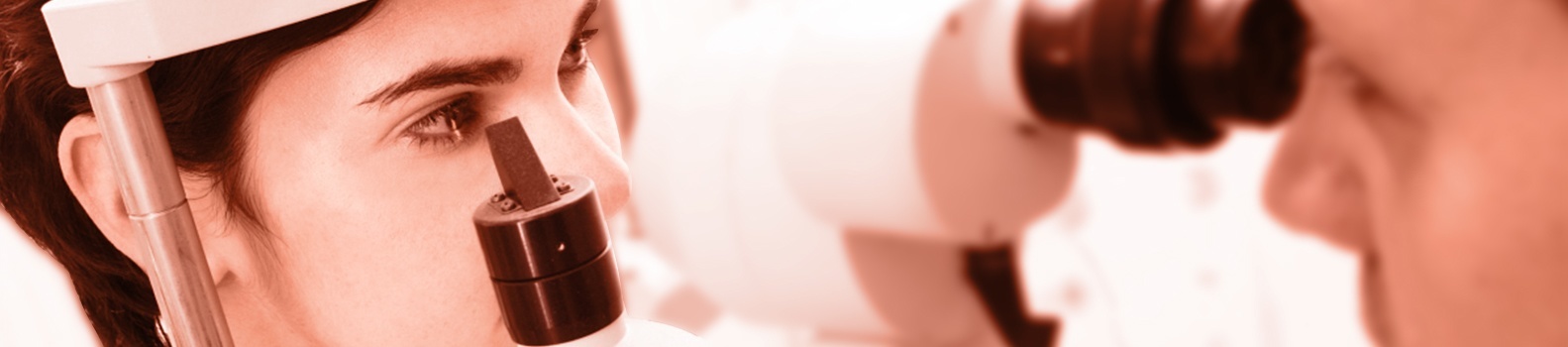
Shroff Eye Hospital is India's First Eye Hospital accredited by the Joint Commission International (USA) since 2006. Shroff Eye is also India's first and only Wavelight Concerto 500 Hz LASIK center. Shroff Eye has stood for excellence in eye care since 1919. A firm commitment to quality is at the heart of all services provided at our centers at Bandra(W) and Marine Drive, Mumbai.
Cornea is the transparent front surface of the eye. Normally, when looking straight on at the eye, you look right through the cornea and see the coloured iris and black pupil of the eye.
The cornea is avascular i.e. contains no blood vessels to nourish or protect it against infection, unlike other tissues in the body. Instead, it receives its nourishment from the tears and aqueous humour which is present in the anterior chamber. It is mainly composed of cells and proteins.
There are a lot of corneal disorders and diseases seen in routine practice. Enumerating below are a few of the common ones:
Refractive errors
If the cornea is flatter than normal or the eye is short, rays of light are focused behind the retina and causes hyperopia or farsightedness where close objects appear blurred.
Astigmatism is a condition in which the uneven curvature of the cornea blurs and distorts both distant and near objects. The cornea is more curved in one direction than in the other. This causes the rays of light to focus on two separate areas of the retina, distorting the visual image.
Refractive errors are usually corrected by eye glasses or contact lenses. Although these are safe and effective methods for treating refractive errors, refractive surgeries are becoming an increasingly popular option.
Allergies
Allergies affecting the eye are relatively common. They are most commonly related to pollen and dust in the air. They are usually immediate or delayed hypersensitivity reactions. Symptoms can include redness, itching, and burning, tearing, stinging and watery discharge. An increasing number of eye allergy cases are related to medications and contact lens wear. Also, animal hair and certain cosmetics, such as mascara, face creams and eyebrow pencil, can cause allergies that affect the eye. Touching or rubbing the eyes after handling nail polish, soaps, or chemicals may cause an allergic reaction. Some people have sensitivity to lip gloss and eye makeup. Mild forms of these allergic disorders are usually restricted to the conjunctiva. However, moderate and severe allergies usually cause affection of the cornea and may lead to decrease vision. These allergies can be treated effectively with topical medications. Hence, every person who gets frequent episodes of redness, watering and itching should consult an ophthalmologist and seek advice.
Conjunctivitis (red / pink eye)
Conjunctiva is a translucent mucous membrane which lines the posterior surface of the eyelids and anterior aspect of the eyeball. A group of diseases that cause swelling, itching, burning and redness of the conjunctiva are termed as conjunctivitis. It is an inflammation of the conjunctiva associated with a discharge which may be watery, mucoid, mucopurulent or purulent. It can spread from one person to another if proper precautions are not taken. It can be caused by a bacterial or viral infection, allergy, environmental irritants, a contact lens product, eye drops or eye ointments. It is usually painless in the beginning and does not affect vision. But in some forms of conjunctivitis, if the infection is severe, it can cause corneal inflammation and loss of vision. Treatment of conjunctivitis is usually with eye drops. However a consultation with an ophthalmologist is a must to make sure that there is no corneal involvement.
Infections
A breach in the normal epithelial surface of the cornea associated with necrosis of surrounding corneal tissue is termed as corneal ulceration. Two main factors are responsible in the production of a corneal ulcer: damage to corneal epithelium and infection of the eroded area. They usually cause pain, redness, watering, discharge, photophobia i.e. intolerance to light and blurred vision. It can be caused by bacteria, fungi, viruses, acanthamoeba and many other organisms. They cause painful inflammation called keratitis. These infections can reduce visual clarity, produce corneal discharge and perhaps erode the cornea. Corneal infections can also lead to corneal scarring, which can impair vision and may require a corneal transplant. As a general rule, the deeper the corneal infection, the more severe the symptoms and complications. It should be noted that corneal infections, although relatively infrequent, are the most serious complication of contact lens wear. Minor corneal infections are commonly treated with anti-bacterial or anti-fungal eye drops. If the problem is severe, it may require surgical intervention. Frequent visits to an eye care professional may be necessary for several months to eliminate the problem.
Dry eye
Dry eye can be caused due to any of the following reasons:
- Aqueous tear deficiency
- Mucin deficiency
- Lipid deficiency
- Impaired eyelid function
- Corneal epithelium abnormalities.
In people with dry eye, the eye produces fewer or less quality tears and is unable to keep its surface lubricated and comfortable. As we age, the eyes usually produce fewer tears. Also, in some cases, the lipid and mucin layers produced by the eye are of such poor quality that tears cannot remain in the eye long enough to keep the eye sufficiently lubricated.The main symptom of dry eye is usually a scratchy or foreign body or sandy feeling as if something is in the eye. Other symptoms may include stinging, irritation, itching, burning or nonspecific ocular discomfort of the eye; episodes of excess tearing that follow periods of very dry sensation; a stringy discharge from the eye; and pain and redness of the eye. Sometimes people with dry eye experience heaviness of the eyelids or blurred, changing, or decreased vision, although loss of vision is uncommon.
Dry eye is more common in women, especially after menopause. Surprisingly, some people with dry eye may have tears that run down their cheeks. This is because the eye may be producing less of the lipid and mucin layers of the tear film, which help keep tears in the eye. When this happens, tears do not stay in the eye long enough to thoroughly moisten it.
Dry eye can occur in climates with dry air, as well as with the use of some drugs, including antihistamines, nasal decongestants, tranquilizers, and anti-depressant drugs. People with dry eye should let their health care providers know all the medications they are taking, since some of them may intensify dry eye symptoms.
People with connective tissue diseases, such as rheumatoid arthritis, can also develop dry eye. It is important to note that dry eye is sometimes a symptom of Sjogren’s syndrome, a disease that attacks the body’s lubricating glands, such as the tear and salivary glands. A complete physical examination may diagnose any underlying diseases.
Artificial tears, which lubricate the eye, are the principal treatment for dry eye. They are available over-the-counter as eye drops. Sterile ointme
Dystrophies
Corneal dystrophies affect vision in widely differing ways. Some cause severe visual impairment, while a few cause no vision problems and are discovered during a routine eye examination. Other dystrophies may cause repeated episodes of pain without leading to permanent loss of vision. The dystrophies are classified according to the anatomic site involved:
Epithelial dystrophies: Epithelial basement membrane dystrophy, Reis Buckler’s dystrophy, Meesman’s dystrophy
Stromal dystrophies: Granular dystrophy, Macular dystrophy, Lattice dystrophy, Schnyder’s crystalline dystrophy
Endothelial dystrophies: Fuch’s endothelial dystrophy, Posterior polymorphous dystrophy, Congenital Hereditary endothelial dystrophy.
Some of these dystrophies do not cause a lot of visual impairment. However most of them require surgery in the form of corneal transplantation for visual restoration.
Keratoconus
Keratoconus is characterized by progressive thinning and ectasia which results in deterioration of the quality of vision and also the quality of life. As the disease begins in young adults, it affects the most productive years of life. So far there has been no effective way to stop the progression of keratoconus. Current methods such as rigid contact lens, & intracorneal ring segments only the refractive error can be corrected without any effect on the progression of keratoconus. It is estimated that eventually 21% of the keratoconus patients require surgical intervention to restore corneal anatomy and eyesight. A new modality of treatment, based on collagen crosslinking with the help of Ultraviolet A (UVA, 365nm) and the photosensitizer riboflavin phosphate has been described which changes the intrinsic biomechanical properties of the cornea, increasing its strength by almost 300%. This increase in corneal strength has shown to arrest the progression of keratoconus in numerous studies all over the world.
Topography guided C3R (Crosslinking) with Topolink
This is done only if you are in early stages and if fit for the same as examined by our doctors as we can additionally also smoothen the shape of the cornea with our laser (PRK), besides strengthening it (C3R).
Accelerated C3R or KXL
The accelerated crosslinking mirrors the traditional crosslinking procedure but differs and benefits patients in 3 ways:
- Reduces cross-linking time from one-hour to a few minutes, adding to patient comfort and experience.
- More importantly, accelerated crosslinking allows thinner corneas to be crosslinked with greater precision, potentially decreasing the risks associated for crosslinking. In effect those patients outside current treatment criteria can be crosslinked and receive the benefits.
- The new Riboflavin solutions can penetrate the epithelium and this procedure is called “Transepithelial Corneal Collagen Cross-linking”.
Pterygium
Pterygia are more common in sunny climates and in the 20-40 age group. Scientists do not know what causes pterygia to develop. However, since people who have pterygia usually have spent a significant time outdoors, many doctors believe ultraviolet (UV) light from the sun may be a factor. In areas where sunlight is strong, wearing protective eyeglasses, sunglasses, and/or hats with brims are suggested. While some studies report a higher prevalence of pterygia in men than in women, this may reflect different rates of exposure to UV light.
A fully developed pterygium has three parts: head, neck and body. Depending upon the progression it may be progressive or regressive pterygium. Progressive pterygium is thick, fleshy and vascular with a few infiltrates in the cornea, in front of the head of the pterygium. Regressive pterygium is thin, atrophic, attenuated with very little vascularity. Ultimately, it becomes membranous but never disappears.
Visual disturbances occur when it encroaches the pupillary area or due to corneal astigmatism induced due to fibrosis in the regressive stage. Occasionally diplopia may occur due to limitation of ocular movements.
Because a pterygium is visible, many people want to have it removed for cosmetic reasons. Other indications for surgery include continued progression threatening to encroach onto the papillary area or diplopia due to differential ocular movements. Pterygium surgery involves excision of the pterygium followed by covering the bare area with a conjunctival autograft either from the same eye or the other one. Recurrence rates are the least when surgery is done with conjunctival autograft technique.
Ocular surface problems
Patients with ocular surface diseases suffer from loss of vision, discomfort, infection, erosions, ulceration, and destruction with scarring of the eye surface. The most common cause of these problems is the imbalance in the neural regulation which leads to an “unstable tear film”. Ocular surface failure manifests in two ways – the first one is the Limbal Stem Cell Deficiency in which the corneal epithelium is replaced by the conjunctival epithelium. In the second one, the corneal or the conjunctival epithelium changes with keratinisation and loss of mucosal epithelial characteristics.
Treatment of these conditions involve ocular surface reconstruction. This involves multidisciplinary approach to restore the normal ocular surface defense so that a stable tear film can be achieved, restore the epithelial stem cells and restore stem cell stromal niche. These treatment strategies combine medical and surgical modalities. Medical treatment includes the use of various topical drops. Surgical modalities include symblepharon release, pannus excision, amniotic membrane transplantation, conjunctival autograft, limbal stem cell transplant etc.
Contact lens related problems
A contact lens wearer who presents with conjunctival irritation and associated symptoms – such as itching, burning or tearing – may be suffering from any one of a number of conditions (such as allergy) of which the lenses may not be the primary cause. In other cases, however, contact lens wear itself is responsible for the condition. The possible conditions which need an ophthalmologist’s opinion for contact lens users include:
- Ocular allergy – tearing, itching, a sensation of ocular pressure, dilatation of conjunctival vessels, swollen eyelids, foreign body sensation
- Dry eye – Irritation, redness, burning/ stinging, itchy eyes, sandy- gritty feeling (foreign body sensation), blurred vision, tearing, contact lens intolerance, increased frequency of blinking, mucous discharge, photophobia (less frequent symptom), symptoms worsen in windy or air-conditioned environments, as day progresses, after prolonged reading, working on computers
- Red eye – there are multitude causes of a red eye in contact lens users. Broadly, they are either infective or noninfective. Amongst the infective causes it can be either conjunctivitis or keratitis. The noninfective causes include: corneal hypoxia, damaged lenses, incomplete blinking, deposits on the lens, poorly fitting lenses and reactions to the lens cleaning solutions. There could be associated, non-contact lens related causes of red eye viz., blepharitis or meibommianitis.
- Corneal problems related to contact lens wear include: corneal abrasions, punctuate keratitis, sterile corneal infiltrates, superior limbic keratoconjunctivitis, neovascularisation and corneal ulcer.
- Giant papillary conjunctivitis: marked by increasing lens awareness, itching, mucous discharge, formation of a coating on the contact lens and development of papillae.
Aphakic /pseudophakic bullous keratopathy
- Regular arrangement of the corneal collagen lamellae
- Lack of blood vessels, lymphatics and cellularity
- Relative state of dehydration which is maintained by the corneal endothelium
- Normal intraocular pressure
Of these, the function of the corneal endothelial cells is of utmost importance. Failure of the corneal endothelial cells to maintain a relative state of dehydration of the cornea results in accumulation of fluid within the cornea known as “corneal edema”. As the corneal stroma swells, there is accumulation of fluid in the subepithelial and intraepithelial spaces which leads to the formation of bullae which causes a lot of discomfort, decreased vision, loss of contrast, glare and photophobia. If these bullae rupture, they cause a lot of pain and foreign body sensation. Bacteria can occasionally cause secondary microbial infection. This is known as Bullous Keratopathy. Most important causes include Fuch’s Endothelial Dystrophy and corneal endothelial trauma. Bullous keratopathy after cataract surgery is called PSEUDOPHAKIC BULLOUS KERATOPATHY (if intraocular lens implant is present) or APHAKIC BULLOUS KERATOPATHY (if intraocular lens implant is absent).
Treatment includes topical dehydrating agents, intra-ocular pressure lowering agents, lubricating agents, bandage soft contact lenses for mild to moderate cases and treatment of any secondary microbial infection. For severe cases, corneal transplantation is usually required.
Till recently, full thickness corneal transplantation was the treatment of choice for these cases. The surgical treatment of corneal endothelial dysfunction has been revolutionized with Descemet’s stripping with endothelial keratoplasty (DSEK). As an alternative to traditional penetrating keratoplasty (PK), DSEK has gained popularity because the selective transplantation of the endothelial layer avoids the potential complications of PK such as wound dehiscence, wound infections, and high postoperative astigmatism. Originally conceived as posterior lamellar keratoplasty by Melles et al, selective endothelial keratoplasty has evolved into deep lamellar with endothelial keratoplasty and now Descemet’s stripping with endothelial keratoplasty (DSEK). In DSEK the recipient Descemet’s membrane and endothelium are stripped and a posterior lamellar graft, or DSEK graft, then is inserted and allowed to unfold with subsequent recipient-to-donor stromal adherence. Adhesion of the DSEK graft allows for eventual deturgescence of the recipient cornea as the donor endothelial cells begin their pump action. Preparation of the posterior lamellar graft, containing the donor posterior stroma, Descemet’s membrane, and endothelium, has been simplified by use of a microkeratome on a corneoscleral button mounted on an artificial chamber. This variant in procedure has been termed Descemet’s stripping automated endothelial keratoplasty (DSAEK).